The internship is halfway over and there have been quite a
few exciting projects and finds in the first 10 weeks. One day, while conserving and cataloging specimens, a furry find was made. Inside a slide, right next to
the specimen, was a larval skin of a beetle! How the larval skin got into the slide and how the beetle got out remains a mystery. This is a great example of why the
conservation work we are doing with old collections is so important.


There have been some really nice scans done of microfossils
as well. Thanks to the Microscopy and Imaging Facility, we have started 3D printing some of the clearest CT scans here at the museum. It has, however, been a bit of
learning curve.


Before you can even think about printing a 3D model, you must first spend time enhancing and editing the 3D image. Here at the American Museum of Natural History we use a program called VGStudio MAX. When a CT scan comes through you hope that the image is clear and that there is little to no sediment infill in the specimen. Even with a perfect scan, it still takes about 2 hours to have a printer-ready specimen. When the scan comes back with infill or "background noise" it can take anywhere from 4-8 hours to edit the image. At first every scan takes 5+ hours and the software can be tricky to get used to. Everyone is different, but I felt comfortable using VGStudio MAX by week 4.

Once you have edited your image, you are ready to start setting up your printing process. One factor we had to look at was which orientation we should print the specimens out for the clearest image. As seen below in the first attempt at printing out AMNH-FI 97614 Hopkinsina magnifica, the bottom model looks a lot nicer and smoother than the one on top.

Once you have edited your image, you are ready to start setting up your printing process. One factor we had to look at was which orientation we should print the specimens out for the clearest image. As seen below in the first attempt at printing out AMNH-FI 97614 Hopkinsina magnifica, the bottom model looks a lot nicer and smoother than the one on top.

Another problem is sizing. Because each
specimen and scan is different, the scaling settings must be customized
depending on the fossil. AMNH-FI 107593 Asterorotalia pulchella printed out too small and had to
be redone so all of the details could be seen properly.

Sometimes everything
can be programmed correctly and the printing still go wrong. There were issues with the printer's plastic filament while printing out AMNH-FI 64208 Anomalina pacoraensis which led to this incomplete 3D
model.


However, after a few mishaps and mistakes, we are starting
to have a nice collection of 3D printed microfossils.

From Top to Bottom: AMNH-FI 19954 Anomalina mantaensis, AMNH-FI 107593 Asterorotalia pulchella, AMNH-FI 97614 Hopkinsina magnifica

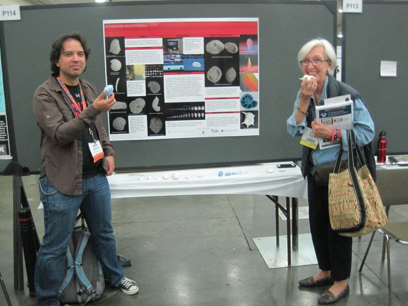
All smiles after printing out this unique foraminifera!