Wednesday, August 19, 2015

Week 8: Then and Now, Final Post

Women, for centuries, have been integral to the science, study, and conservation of invertebrate paleontology/zoology. However, until recently, they were rarely acknowledged for their work and incredible contributions to their respective fields. Female trailblazers of natural history and science include Mary Anning, Libbie Hyman, Mary Leakey, Patricia Kelley, and Susan Kidwell, to name only a few. All the interns would like to acknowledge and put the spotlight on two of today's trailblazers, Ellen Thomas and Bushra Hussaini, who have led this microfossil preservation and conservation project at the AMNH.


Morgan was thrilled to have worked with Ellen for eight weeks
Ellen Thomas

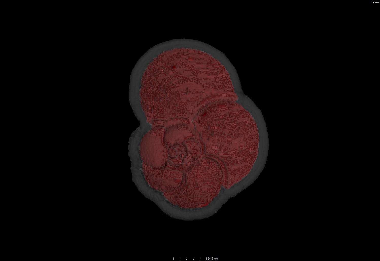
Early on, Ellen was interested in the sciences, but thought about chemistry and biology, not geology or paleontology. Then she went to Utrecht University (The Netherlands) information days in 1968 to look at options in the sciences, and was told that women do not study geology. That was the fire and drive for her to decide to become a geologist. She never looked back, earning a bachelor’s and master’s degree in Geology, then a Ph.D. studying benthic foraminifera. For the next 35 years or so, Ellen has studied microfossils to make major advances in the field of paleoceanography, which eventually led her to the AMNH. To us interns, Ellen is the "foram expert" and we take every Tuesday to absorb her knowledge and admire her passion for the historic foraminifera and ostracod collections for the benefit of future scientists.


Sam and Bushra enjoying the spectacular view over Central Park West.
Bushra Hussaini

Travelling with her father in the northwestern parts of Pakistan developed Bushra's love for mountains and nature. However, it was not until she got involved with rocks, minerals, and mapping at the University of Karachi did she passionately pursue her Bachelor's and Master's in Earth and Environmental Sciences while being the only woman to graduate in the top 10 students of her class. Immigrating to the United States in 1984, Bushra found a job working as a research assistant at Lamont Doherty Geological Observatory. She eventually ended up working at the AMNH as a Senior Scientific Assistant and has been managing the Fossil Invertebrate collections since 1997. We all consider Bushra to be kindhearted, patient, understanding, intelligent, and witty. She has shown all the interns what it means to be a respected professional and mentor.


Today, four more female trailblazers have made their mark at the AMNH. Lindsay, Morgan, Brittney, and Claudia each have bright futures and inevitably will rise to the top of their respected field of study. It has been a pleasure to work with all of them.

Acknowledged invertebrate paleontologists on the steps of the AMNH during the early 1900s and today's stars making their mark 100 years later.

Week 6: More than Microfossils

Working on collections that haven't been looked at in nearly 100 hundred years always comes with challenges, and sometimes even some creepy crawlies. The collection previously used wooden boxes to store slides containing micro-fossils and these boxes allowed, and welcomed, some unwanted and unexpected critters of the arthropod variety to set up camp next to our micro-fossils.

This was found in a box of miscellaneous slides by Sam while rehousing specimens. The intruder, identified as a carpet beetle, was found alive scampering away across the table. 


This creepy crawly was found while Brittney was rehousing slides. There were two of these mysterious web-like cocoons on one slide, and several throughout a study collection. It appears to have a leg, possibly molted, of unknown origin dangling from the cocoon.   


This ostracod, Cythereis asperima, from the Schmidt 1939 collection was not only fastened onto the slide by glue, but cob webs too! There were nearly ten slides that exhibited evidence of arachnid presence.  


These intruders of creepy crawlers frequent museum collections, especially if the collection isn't properly cared for. Finding these on our slides and in our collection reiterates the importance of this project. Specimen preservation, and collection managing have changed significantly since these microfossils were first accessioned. For example, the reason why so many ostracods of the Schmidt, 1939 collection were covered with spider webs is because the glue used to secure the specimen to the slide is organic and apparently attracted spiders. In order to remedy this widely accepted use of organic glue in museum collections, the specimens must be cleaned and moved to fresh slides, and the old slide is discarded to prohibit any more unwanted guests.  

As time continues our methods and processes of collection management will continue to advance, allowing museum collections such as the AMNH microfossils, to be preserved and safe for years to come.  

Tuesday, August 11, 2015

Week 5: Day at the Museum


We've had a lot of adventures here at the museum so far! All of the interns have been learning and developing new skills and tackling some pretty intimidating challenges.

Lindsay calmly gets her work done, and Sam never loses his cool. Claudia handles the pressure worst of all.

For example, Lindsay, Sam and Claudia were given a collection of Permian fusulinids from the late Paleozoic. This collection included over 500 slides!

The collection needed re-housing, entry into the database and lots of imaging! Everyone gained a few new grey hairs, but were very proud of the finished products.

A light microscope image of the holotype, Pseudoschwagerina vilcanotensis.
Another challenge for the interns is CT Scanning. This amazing technology allows us to create 3D images of our microfossils and see the outer shell and internal structure at great resolution.

Processing these files can be slow, but Morgan finds plenty of coffee helps get the job done.


However, you can't beat that amazing view!


 Top: Bulimina rectospinata Bottom: Globorotalia menardi

The work that we are doing at AMNH is very important for the preservation of the collections and for scientists interested in microfossils around the world. And if stress ever builds up too much, we can always take advantage of our employee discount at the museum store!

Swag modeled by Brittany: AMNH fleece jacket and T. rex-embroidered AMNH scarf! 

Friday, July 24, 2015

Week 4: Half Way There Captions


Morgan can't get enough processing of CT scans in the Microscopy and Imaging Facility.


                     Brittany thrilled about curating invertebrates in the Vertebrate Paleontology department.


Claudia has never seen so many hard rocks as she did in the Planetary Science division.


                                           Sam trying to hide behind the 500+ thin sections of the Robert's collection.


Lindsay expressing her disbelief and disappointment at the frozen tissue lab.

                    Bushra showing the interns the magnificence of the Invertebrate Paleontology collection.


Claudia enjoying her BLTF while out in Central Park for lunch.


Week 3: Is Beauty Really Only Test Deep?


  (http://www.explainxkcd.com/wiki/index.php/460:_Paleontology acessed on 7/14/15)


There is no denying that interest in macrofossils partially stems from how aesthetically pleasing (or interesting) they are. However, microfossils are incredibly easy on the eyes as well... as long as the light on your microscope isn't on a blindingly bright setting. This week we will be highlighting some of our most beautiful Foraminifera and Ostracods, as well as the Computed Tomography scanning that is used when we want to explore internal structures. Because beauty isn't only test deep, right? 


Elphidium fax fax, a paratype associated with David Nicol's publication titled "New West American Species of the Foraminiferal Genus Elphidium", published in the Journal of Paleontology, 1942.


Elphidium fax barbarense, a paratype associated with David Nicol's publication titled "New West American Species of the Foraminiferal Genus Elphidium", published in the Journal of Paleontology, 1942.


Tuberitina bulbacea, a holotype associated with Galloway and Harlton's publication titled "Some Pennsylvanian Foraminifera of Oklahoma, With Special Refrence to the Genus Orobias", published in the Journal of Paleontology, 1928.


Bairdoppilata pondera, our beautiful Ostracod this week, is a holotype associated with Jenning's publication "A Microfauna from the Monmouth and Basal Rancocas Groups of New Jersey", published in the Bulletins of American Paleontology, 1936.


Elphidium cynicalisa holotype associated with Jenning's publication "A Microfauna from the Monmouth and Basal Rancocas Groups of New Jersey", published in the Bulletins of American Paleontology, 1936.


Cibicides burlingtonensis, a holotype associated with Jenning's publication "A Microfauna from the Monmouth and Basal Rancocas Groups of New Jersey", published in the Bulletins of American Paleontology, 1936.


Aren't they pretty? Give them a round of applause! Or at least try..

 
(https://pbs.twimg.com/media/B9nWTuCCcAAoStA.jpg acessed on 7/14/15)


So what happens when we want to see beyond the external structure of a specimen? Luckily, here at the museum we are equipped with a CT scanning machine, or "Computed Tomography", which translates to the imaging of objects by sections with the use of a penetrating wave. A CT scanner bombards an object with x-rays as the object rotates around a central axis. As the object rotates, thousands of images are collected from all angles, which are then compiled to create a 3 dimensional reconstruction of the object. This allows us to see beyond the surface of a specimen! 

Below is an image of a project that intern Lindsay Walker is working on. She is finishing up the reconstruction of  Foraminifera Guadryina from Diamond 1928, an unpublished master's thesis. 


You can see in the lower right corner of the screen capture that the scanning of the image allows us to see the different chambers of the organism, all without having to do any physical opening of the specimen. 

Below is intern Claudia Deeg completing the reconstruction of Epistomina flinti, a holotype specimen from Galloway and Wissler's publication titled "Pleistocene Foraminfera From the Lomita Quarry, Palos Verdes Hills, California", published in the Journal of Paleontology, 1927.


Last but not least, intern Morgan Black completed a reconstruction of holotype specimen Cythereis tremoidia, from  R.A.M Schmidt's Columbia University Master's Dissertation, titled "Miocene Ostracoda from Yorktown Formation Virginia", published in 1939. Below is a video of the final product. 


Not only are these microfossils useful, but they sure are good-lookin'!

Tuesday, July 7, 2015

Week 2: What's in the box?

By Lindsay Walker

This year the micropaleontology interns will dedicate most of their time to cleaning up unfinished projects from the previous two summers, working through the remaining quota of images and CT scans, and, finally, wrapping up with quality control checks. At the AMNH, each microfossil collection typically corresponds to a paper (published or unpublished), usually written in the mid-20th century or earlier. Part of what makes our work interesting is that every collection is different and, consequently, presents its own curation challenges. Below are descriptions of some of the projects we’ve been working on during our first two week at the museum.


~~~~~~~~~~~~~~~~~~~
Dorr? (Year?)
No publication
Collection of foraminifera similar to those gathered by the H.M.S. Challenger Expedition (1873-1876)


Not every microfossil collection can be clearly associated with a publication, and sometimes specimen slides contain misleading information. For example, this week Brittney was assigned a collection that appeared to have been collected by the famous H.M.S. Challenger expedition, which led one of the earliest attempts to document marine life across the Atlantic, Pacific, and Indian Oceans. The resulting monograph was authored by Henry Brady (1884), a pioneer in the study of foraminifera, and is still widely used today for its high-quality illustrations. Needless to say, this collection was a potentially exciting find. Only after consulting our resident foram expert was it determined that the collection was actually made by an unknown collector--possibly "Dorr"--who may have revisited some of the Challenger's original collecting localities. Brittney has been matching specimens from this collection to their locality information, including forams from Fiji and Tonga (27 slides) and Sable Island (unquantified # of slides).

Route of the H.M.S. Challenger Expedition
Brady’s monograph contains detailed illustrations relative to most other publications the interns are working with (compare to Diamond 1928, shown below).


~~~~~~~~~~~~~~~~~~~
Coryell & Cuskley (1934)
Some ostracodes from the “White Mound” section of the Haragan Shale, 
Murray County, Oklahoma
Description of lower Devonian ostracods published in American Museum Novitates 

Morgan’s first collection did not contain foraminifera, but was a large collection of ostracods collected by H. N. Coryell of Columbia University. The collection was partially completed by a former intern, but 40 slides remained to be curated, rehoused, and databased. Morgan was responsible for completing these tasks, as well as imaging one paratype to compliment the 16 previously imaged holotypes.

Paratype specimen Condacypris bindoa (AMNH-FI 103075)
Image by Morgan Black

~~~~~~~~~~~~~~~~~~~
Diamond (1928)
Oligocene Foraminifera from Palma Real, Vera Cruz, Mexico
Unpublished thesis by Columbia University master’s student


The Diamond collection was Lindsay’s first project. It contains 45 slides of foraminifera, several of which may contain new species. This collection was completely uncurated when Lindsay received it, meaning she had to catalog, database, and rehouse every specimen present, as well as document any missing slides or lost specimens. All potential new species were imaged, and one of these specimens, Gaudryina n. sp., was added to the CT scanning queue. Because the results of this master’s thesis were unpublished, it is likely that these specimens have remained untouched in the museum’s cabinets for nearly a century.
Comparison of hand-illustrated specimens and a digitally imaged specimen from the Diamond thesis
Image at right by Lindsay Walker

~~~~~~~~~~~~~~~~~~~
Kling (1960)
Permian fusilinids from Guatemala
Analysis of fusilinid foraminifera published in Journal of Paleontology


As the invert paleo interns learned from Ellen Thomas this week, fusilinids comprise a group of foraminifera that disappeared along with >90% of other marine species during the end-Permian mass extinction event (~250 Ma). These forams differ from other foraminifera in that they are much larger (visible to the unaided eye) and their tests (shells) are structurally more complex. This week Sam imaged and databased 40 petrographic thin sections from this collection of fusilinid foraminifera. That this collection contains only thin sections is unusual; most of the other AMNH microfossil collections consist of specimens glued to cardboard slides. This meant Sam had to image every slide in this collection as opposed to type specimens only, as is the standard protocol for collections mounted on cardboard slides.

Thin sections showing the cross-sectional views of fusilinid foraminifera
Images by Sam Martin

Week 1: Introducing the 2015 Intern Team

On Monday, June 22, the final group of paleontology interns arrived at the AMNH to help with the ongoing microfossil conservation and imaging project started in 2013. For our first week, we’ll be introducing ourselves as you’ll be hearing more about us in future blog entries.

Left to right: Claudia, Lindsay, Morgan (front), Brittney, and Sam will be helping complete the microfossil conservation and imaging project started in 2013. They hope the diversity of their past experiences will help them meet this challenge under the guidance of Bushra Hussaini, Senior Scientific Assistant in the Division of Paleontology.

Morgan Black is a current undergraduate student enrolled in a B.S. Geology degree program at Morehead State University. She has an emphasis in biogeosciences and is currently a palynology research fellow under Dr. Jennifer O'Keefe. Morgan is currently researching a set of fluvial/lacustrine deposits in eastern Kentucky and plans to present preliminary paleoecological data at the annual GSA meeting in Baltimore this fall. She was the only winner in the United States of the American Association of Stratigraphic Palynologists undergraduate award in the 2014-2015 academic year. She is very excited to be here at AMNH gaining more experience in the field of micropaleontology, along with databasing and computed tomography, and hopes to bring back much of what she learns here in the museum to her university.

Claudia Deeg, like the other interns, is not a New York City native, but is originally from Philadelphia. She is an upcoming junior at Smith College where she is working toward her undergraduate degree in biology with a minor in geology. At Smith, Claudia has been studying foraminifera spanning the Cretaceous-Paleogene extinction event under Dr. Sara Pruss. This is her first time working in a museum and Claudia is particularly looking forward to learning more about the technology used to image microfossils.

Sam Martin is a returning intern from 2013. Since then, he has graduated from Southern Illinois University with a honors B.S. Geology degree. He is now going into his second year of graduate school at East Carolina University where he is pursuing a master’s degree in Geological Sciences. His research as a graduate student continues to focus on foraminifera. His working thesis, entitled Distribution and taxonomy of modern benthic foraminifera of the Sunda Shelf (South China Sea) off peninsular Malaysia will, for the first time, document the foraminiferal assemblages and diversity present in the shallow waters of the western South China Sea. He is excited to take a break from his own thesis and help finish the microfossil conservation and imaging project he helped start two summers ago.

Brittney Oleniacz is a current master's student at the University of Kansas in Museum Studies with an emphasis in natural history. Her thesis focuses on the public understanding of natural history museums and the research they produce. During the academic year, she is employed by the KU Biodiversity Institute on a digitization project in the invertebrate paleontology collection. She is interested in understanding microfossil taxonomy, an unfamiliar group to her, and gaining more experience in digitizing museum collections, making them accessible worldwide.

Lindsay Walker is a recent graduate of the Museum and Field Studies M.S. program at the University of Colorado-Boulder. She worked with microfossils and fossil insects for her undergraduate and master’s theses, respectively, and her graduate program emphasized natural history collections management. Lindsay is looking forward to learning how to navigate the challenges of curating and conserving historic research collections. Because imaging has become an increasingly important facet of collections management, she is also excited to work with CT scan data, among other imaging techniques, while at the AMNH.